In the diagnosing section of any medical facility, we find a big variety of equipment. This is used to get medical images of the patient for diagnostic purposes. It may include A wide variety of equipment used to produce patient medical images. This includes x-rays, MRIs, ultrasounds, optical imaging, etc.
With time there is a distinct development and progress in this medical field. Some of the latest technologies in this field are beneficial to maintain patient data and storing patient images. Others are to present the medical images in a more precise way to help an accurate diagnosis of the issue.
What are the Prominent Advancements in the Medical Imaging Technology?
Medical imaging is the most important part of any medical facility center. It helps a lot to save millions of lives each year. From cancer and an appendix infection to heart attacks and strokes, it is the method of choice for doctors when it comes to identifying their patients’ conditions. However, the healthcare sector is constantly striving to bring patients cutting-edge innovations. This is why many groups put money into developing better medical imaging technologies.
Let’s take a look at the most recent developments in medical imaging technology.
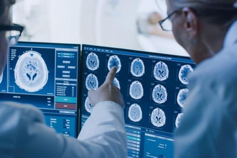
1. Artificial Intelligence
Artificial intelligence (AI) is basically the ability of machines to simulate human intelligence. This is especially true of mental processes like learning and solving problems. AI can be taught to analyze medical images for abnormalities in human tissue, which will help with disease diagnosis and treatment tracking. Radiologists can benefit from artificial intelligence in three ways. Artificial intelligence is capable of searching through massive amounts of patient data and images at lightning speeds. So the results can accelerate. Second, AI can be taught to spot inconsistencies that human eyes would miss.
The precision of diagnoses may benefit from this. Finally, AI can be used to find previous imaging scans from the EMR and compare them to the most recent scan results for a patient. The electronic medical record (EMR) can be retrieved in its entirety, including any relevant medical history, to help with the diagnosis.
There have been several successful attempts to integrate AI into imaging systems by different companies, but as of yet, none of these systems are available for widespread commercial use. Viz is an example of medical imaging software that incorporates AI to better detect and diagnose large vessel obstructions in patients. (LVOs). The software can search for LVOs in multiple image databases from different hospitals. The software can notify both the stroke specialist and the primary care physician if an LVO is detected, increasing the likelihood that the patient will receive timely treatment. Improving outcomes and decreasing the financial burden on the healthcare system is the result of a time-sensitive disease like stroke.
2. Augmented Reality
Augmented reality (AR) and virtual reality (VR) are paving their way in the current healthcare system. It has many applications. Radiological scans of a patient are used in pre-operative planning by surgeons. With the advent of 3D imaging, doctors now have a clearer picture of the patient’s anatomical makeup than ever before. However, determining the precise location of an area in real-time has proven difficult during surgical procedures. The most effective remedy has been fluoroscopy assistance during surgical procedures.
Surgeons can use augmented reality technology to view medical images on AR headsets while operating. The OpenSight AR system from Novarad is an FDA-approved augmented reality (AR) solution for pre-surgical planning. Patients’ radiation exposure is effectively lowered, and surgeons’ preoperative planning is enhanced.
3. Picture Archiving and Communications System (PACS)
A radiology picture archiving and communication system is one type of medical imaging management solution. PACS stands for “Picture Archiving and Communications System,” a technological solution developed to alleviate the burden of managing the massive quantity of medical images generated by the radiology department.
There are many ways in which a PACS can be used to facilitate hospital-wide image management in medical imaging. Surgical and cardiology suite images, as well as emergency department images, etc., can be linked to a nationwide network of hospitals and imaging centers. Because of cooperation, tasks can be completed more quickly, saving a great deal of time. In the long run, this will help doctors and nurses become more efficient, which will improve patient care and management.
4. Digital Mammography
It is extremely beneficial in terms of diagnosing breast cancer. Digital mammography offers an even higher level of accuracy than is required in film and X-rays. These were older methods that produced the same results. Digital mammograms are more effective than traditional ones. This is especially true for women with thick tissue in their breasts, women nearing the the menopause age, women who are premenopausal, and those below 50 years of age.
The process of creating these images along the breast is called digital breast tomosynthesis (DBT). The radiologists’ ability to see the lesion and detect cancer at an early stage is greatly enhanced by the ability to view each tissue separately, rather than by combining two projections of images. As a result, there will be fewer false positives and recalled patients. Finally, eliminating radiation from mammography without sacrificing image quality is made possible by combining digital mammography with ultrasound and MRI.
5. Open and Portable MRIs
Going through an MRI scan is not something entertaining. Using a “large magnet, radio waves, and a computer,” magnetic resonance imaging (MRI) can produce “a detailed, cross-sectional image of internal organs and structures.” In most cases, the machines will be in the shape of a long tube with a table inside. Patients have often felt anxious during traditional MRIs, particularly those who suffer from claustrophobia. No one would choose to be in an environment with constant whirring noises and a sense of confinement. Open MRI machines are now available, which are less confining and have larger side windows. Modern scanners are much more forgiving of body size than their predecessors.
Future Trends in Medical Imaging
The next development in imaging is 4D medical imaging. This cutting-edge imaging technique significantly improves the rate and precision of image analysis. In order to better observe and analyze motion and variation, 4D medical imaging combines three-dimensional (3D) images with time.
Conclusion
The forefathers of medical imaging technologies probably had no idea that their inventions would evolve into what we have today. Improvements in imaging technology have made way for faster and more precise diagnoses in modern medicine. These days, radiologists not only perform diagnostic imaging but also administer radiation therapy, supervise interventional procedures, and train other medical imaging professionals.